animal cell under microscope labeled
You will find two main parts in hair a cylindrical shaft and a terminal hair follicle. Make a wet or dry mount with a coverslip.
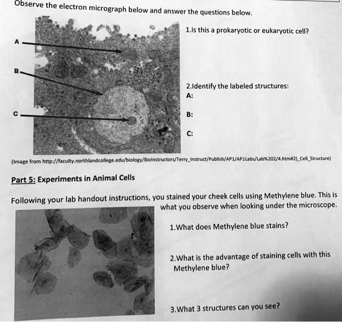
Solved Observe The Electron Micrograph Below And Answer The Questions Below 1 Is This A Prokaryotic Or Eukaryotic Cell 2 Identify The Labeled Structures Image From Htto Faculty Northlandcollegeedu Blology Biolnstructors Terryinstruct Publish Apl
Hair under microscope.

. Part of its innovative design is protected by intellectual property IP laws. This cellular compartment is found only in those bacteria that have both an outer membrane and plasma membrane eg. A nucleus or a cell wall can be seen more clearly by using different stains.
The unique entity identifier used in SAMgov has changed. This neuron possesses the cell body an axon and several short branched dendrites. In contrast the nucleus of a plant cell is located on one side of the cells.
Welcome to Patent Public Search. Every cell has a cell membrane whether it be a plant or animal. The multipolar motor neuron under a microscope shows the typical features.
From Latin nucleus or nuculeus meaning kernel or seed is a membrane-bound organelle found in eukaryotic cellsEukaryotic cells usually have a single nucleus but a few cell types such as mammalian red blood cells have no nuclei and a few others including osteoclasts have manyThe main structures making up the nucleus are the. This subpopulation corresponds to neuronal and neural-progenitor-like tumor cell states as defined by single-cell transcriptomics both in mouse models and in the human disease. Reporting on information technology technology and business news.
Composed of peptidoglycan polysaccharides protein the cell wall maintains the overall shape of a. The cell nucleus pl. A cell membrane is a division between the outside environment and the inside protoplasm of the cell.
February 5th 2021 - Updated October 1st 2021 March 12th 2022. A MESSAGE FROM QUALCOMM Every great tech product that you rely on each day from the smartphone in your pocket to your music streaming service and navigational system in the car shares one important thing. The neurons in the ventral horn of the spinal cord and cerebral cortex are the best examples of multipolar motor neurons.
Immunofluorescence utilizes fluorescent-labeled antibodies to detect specific target antigens. How to see the cell nucleus under a microscope. In chemistry an inorganic compound is typically a chemical compound that lacks carbonhydrogen bonds that is a compound that is not an organic compound.
Structure Parts Functions Labeled Diagram. The Amoeba Sisters walk you through the reason for mitosis with mnemonics for prophase metaphase anaphase and telophase. In an animal cell the nucleus is typically located in the central region of the cell.
To ensure that all Wikipedia content is verifiable Wikipedia provides a means for anyone to question an uncited claimIf your work has been tagged please provide a reliable source for the statement and discuss if needed. Gram negative bacteriaIn the space are enzymes and other proteins that help digest and move nutrients into the cell. You will find the cell base on the basement membrane and the apex in contact with the lumen.
The possible existence of unseen microbial life was suspected from ancient times such as in Jain scriptures from sixth century BC India. In 2015 AquAdvantage salmon became the first genetically modified animal to be approved for food use. The Patent Public Search tool is a new web-based patent search application that will replace internal legacy search tools PubEast and PubWest and external legacy search tools PatFT and AppFT.
You can add a citation by selecting from the drop-down menu at the top of the editing boxIn markup you can add a citation manually using ref tags. Because of its specificity. The tiny iridescent Barrens topminnow spent more than 40 years in endangered species limbo under on-and-off review but never officially listed as endangered October 21 Report.
Most of the cells are microscopic hence they can only be seen under a microscope in order to study their anatomy. A karyotype is a preparation of the complete set of metaphase chromosomes in the cells of a species or in an individual organism sorted by length centromere location and other features and for a test that detects this complement or counts the number of chromosomes. The first genetically modified animal to be commercialized was the GloFish a Zebra fish with a fluorescent gene added that allows it to glow in the dark under ultraviolet light.
See how a generalized structure of an animal cell. The scientific study of microorganisms began with their observation under the microscope in the 1670s by Anton. The study of inorganic compounds is a subfield of chemistry known as inorganic chemistry.
The cylindrical shaft of the hair under a microscope shows three layers medulla cortex and cuticle of keratinized cells. Microscope cell staining is a technique used to improve the visibility of cells and cell parts under a microscope. On April 4 2022 the unique entity identifier used across the federal government changed from the DUNS Number to the Unique Entity ID generated by SAMgov.
Cell is a tiny structure and functional unit of a living organism containing various parts known as organelles. Differentiate between a condenser and an Abbe condenser. The Unique Entity ID is a 12-character alphanumeric ID assigned to an entity by SAMgov.
Simple columnar epithelium under a microscope The simple columnar epithelium under a microscope consists of a single layer of taller than wide cells. Animal Cell- Definition Structure Parts Functions Labeled Diagram Prokaryotes vs Eukaryotes- Definition 47 Differences Structure Examples Amazing 27 Things Under The Microscope With Diagrams. A microorganism or microbe is an organism of microscopic size which may exist in its single-celled form or as a colony of cells.
They are found under the stage next to the diaphragm of the microscope. Karyotyping is the process by which a karyotype is prepared from photographs of chromosomes in order to. Learn the structure of animal cell and plant cell under light microscope.
Tumor cell invasion resembled neuronal migration mechanisms and adopted a Lévy-like movement pattern of probing the environment. These simple columnar epithelia look closely packed and slender columns shaped. Condensers are lenses that are used to collect and focus light from the illuminator into the specimen.
Inorganic compounds comprise most of the Earths crust although the compositions of the deep mantle. Amazing 27 Things Under The Microscope With Diagrams. News for Hardware software networking and Internet media.
It was released to the US market in 2003. Animal cell definition with cell size and shape. So hair is an epidermal down growth embedded into the dermis or hypodermis of the animals skin.
Prokaryotes vs Eukaryotes- Definition. Cell organelles structure and parts. They play a major role in ensuring clear sharp images are produced with a high magnification of 400X and above.
Iodine crystal violet and methylene blue are examples of simple stains. Multipolar motor neuron under microscope. Elon Musk plans.

81 481 Plant Cell Stock Photos Pictures Royalty Free Images Istock

Calendar

Draw A Diagram Of Animal Cell And Label Any Three Parts Which Differentiate It From Plant Cell Brainly In
Plant Cell Definition Structure Parts Functions Labeled Diagram

Cell Micrographs Bioninja
Amazing 27 Things Under The Microscope With Diagrams

Gce Cie Biology Animal And Plant Cell Structures And

1 2 Difference Between Plant And Animal Cells Cells As The Basic Units Of Life Siyavula

Lab Manual Exercise 1
Sperm Under Microscope With Labeled Diagram Anatomylearner The Place To Learn Veterinary Anatomy Online

1 2 Difference Between Plant And Animal Cells Cells As The Basic Units Of Life Siyavula

Animal And Plant Cells Microscope Slide Set Microscope Sample Slides Amazon Com Industrial Scientific
Draw And Label The Animal Cell

Cellular Biology And Microscopy Ppt Download Cell Organelles Animal Cell Structure Organelles
Amazing 27 Things Under The Microscope With Diagrams

Given Below Is A Diagrammatic Sketch Of Electron Microscopic View Of An Animal Cell A Label The Brainly In
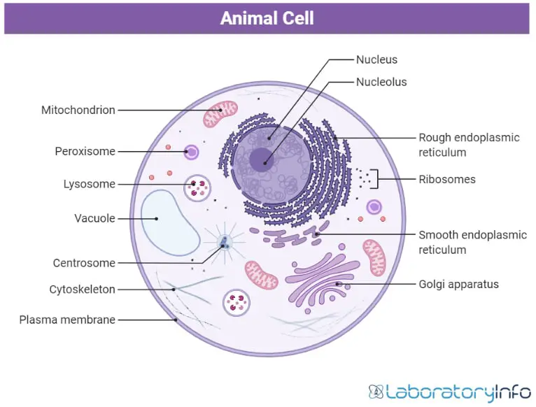
Detailed Guide On Animal Cell And Its Parts With Labelled Diagrams Laboratoryinfo Com
What Organelles Would Be Visible In A Cheek Cell Why Quora
What Is A Diagram Of A Plant And Animal Cell Under An Electron Microscope Quora